


The knee gets blamed for problems that originated several joints away. Of all the upstream sources, the ankle is the most commonly overlooked. Limited ankle dorsiflexion — the motion that brings the front of the foot toward the shin — forces compensations through the entire lower body. The knee, hip, and lower back end up doing work that should have been distributed across the system.
The cascade is consistent. When the ankle can't dorsiflex enough during a squat, the knee either tracks inward (valgus) or the foot rolls inward (pronation) or the lower back arches to maintain forward lean. None of these are designed for repeated loading. Over months and years, the patterns produce overuse injuries that get treated at the symptom site without ever addressing the source.
This piece walks through how to assess your ankle mobility, why it limits the rest of the kinetic chain, and what to do about it.
What Ankle Mobility Actually Means
Most "ankle mobility" discussion focuses on dorsiflexion specifically — the motion that brings the foot upward (front of foot toward shin) with the heel staying down. This motion happens primarily at the talocrural joint, where the tibia and talus articulate.
Functional dorsiflexion requires:
- The talus to glide posteriorly under the tibia
- The calf muscles (gastrocnemius and soleus) to lengthen
- The Achilles tendon to extend
- The joint capsule to allow the motion
- The deeper plantar flexors (tibialis posterior) to allow lengthening
Limitations can come from any of these. Calf shortness is the most common in non-athletes. Joint capsule tightness is common after old sprains. Bone-shape variations affect some people structurally.
Other ankle motions matter for foot mechanics — plantarflexion (pointing the foot down), inversion (sole toward midline), eversion (sole away from midline), and the complex motions of the subtalar joint. But for the lower-body kinetic chain in squatting, running, and most loaded movement, dorsiflexion is the dominant variable.
The Knee-to-Wall Test
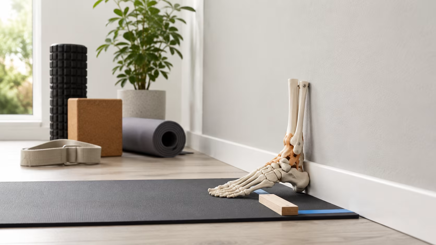
A reliable, replicable test for dorsiflexion:
- Face a wall.
- Place one foot perpendicular to the wall, with the big toe approximately 10 cm from the wall.
- Without lifting the heel, drive the knee forward to touch the wall.
- If the knee touches without heel lift, move the foot another 1-2 cm back and repeat.
- Find the maximum distance at which the knee can touch the wall with heel down.
- Measure that distance. Test both sides.
Interpretation:
- More than 12 cm: above-average dorsiflexion. Adequate for nearly any movement.
- 10-12 cm: normal range. Adequate for most squatting and running.
- 8-10 cm: mild restriction. May cause compensation during deep squatting; usually adequate for running.
- 6-8 cm: moderate restriction. Will affect squat depth, may produce knee pain during running, often compensated in lower-back arching during squatting.
- Less than 6 cm: significant restriction. Substantial compensation in any loaded lower-body movement.
Asymmetry is common and matters. A 12 cm side and an 8 cm side will produce uneven loading patterns. Work the limited side specifically.
The test is sensitive enough that daily mobility work usually shows improvement of 1-2 cm within 2-3 weeks. The metric makes progress measurable, which helps adherence.
Why Limitations Develop
Several common contributors:
Old ankle sprains
The single largest predictor of adult ankle stiffness. A 2013 study by Doherty and colleagues found that even one significant ankle sprain produced lasting dorsiflexion deficits in over half of those affected. The residual capsular tightness from incomplete rehab persists for years.
Calf shortening from chronic plantarflexed position
High-heeled shoes (even modest heels), lots of sitting (which leaves the calf in a slightly shortened position), and lack of intentional calf stretching all contribute. The gastrocnemius and soleus shorten over decades.
Limited use of full range
The body conserves what it doesn't use. Most adults don't routinely squat deep, walk barefoot on uneven surfaces, or load the ankle through full available range. The mobility decays.
Anterior talar shift
With repetitive impact (running, jumping) without adequate posterior glide, the talus can sit slightly forward in the joint. This restricts the posterior glide that dorsiflexion requires.
Footwear with significant heel elevation
Most running shoes have 8-12 mm of heel-to-toe drop. Years of this position contributes to calf shortening. Minimalist footwear advocates argue this is a major modern issue; the evidence is mixed but the mechanism is plausible.
Bone shape
Some people are anatomically less mobile. The tibia, talus, and fibula architecture varies, and ankle range is partly structural.
Specific medical conditions
Equinus contracture, certain neurological conditions, and post-surgical scarring all produce specific limitations beyond what general mobility work addresses.
For most desk-bound adults, the dominant contributor is sustained reduced range usage combined with calf shortening. Both respond well to consistent work.
What Compensation Looks Like
When the ankle can't dorsiflex enough during a movement, several compensation patterns emerge:
Heel lift during squat
The heel comes off the floor at the bottom of the squat. Shifts the load forward, increases knee shear, reduces glute activation.
Knee valgus
Knees collapse inward. Tracking inside the foot rather than over it. Common cause of patellofemoral pain syndrome.
Foot pronation
Foot rolls inward, arch flattens. The body finds dorsiflexion-like motion through the arch instead of the ankle joint. Stresses the medial knee and the medial arch.
Forward trunk lean
The torso pitches forward to keep the bar over mid-foot. Stresses the lower back. Common in long-femur lifters but exaggerated by ankle restriction.
Reduced squat depth
Squats stop at parallel or above. Less hip and knee range trained.
Asymmetric loading
The more mobile side absorbs more depth; the less mobile side bears less load. Produces asymmetric strength development and risks.
Running gait alterations
Overstriding to compensate for reduced ankle ROM during midstance. Increased impact loading. Knee and shin issues follow.
The pattern: the ankle's failure to do its job doesn't cause the ankle to hurt. It causes the next joint up to do compensatory work and hurt. Knee pain is the most common downstream consequence; lower back pain runs a close second.
Mobility Drills That Work
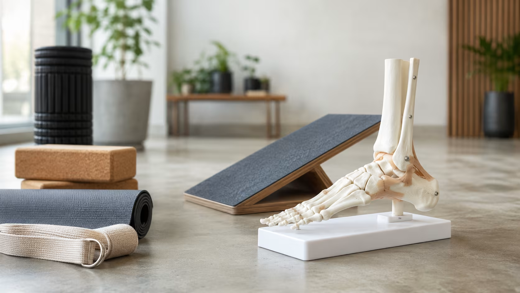
A baseline mobility set for ankles:
Knee-to-wall mobilization
The test position used as the drill. Face a wall, foot 10-12 cm from wall (or wherever your max is). Drive knee forward to touch wall, return. 10-15 reps each side. Progress by moving the foot back over weeks.
Loaded ankle mobilization with band
Loop a thick resistance band around the ankle just above the joint, anchored behind. Step forward into a lunge position. The band pulls the talus posteriorly. Drive the knee forward over the foot through the band's resistance. 10-15 reps. This drill addresses the posterior glide that simple stretching often doesn't reach.
Calf raises through full range
Standing on edge of step, drop heels below toe level, then raise as high as possible. Builds strength through full ankle ROM. 3 sets of 12-15 with bodyweight; progress to single-leg.
Deep squat hold
Bottom of a deep squat position, held for time. 30 seconds to 2 minutes. Loads dorsiflexion at maximum range. Hold a support if needed early. Multiple times per day if possible.
Soleus stretch with bent knee
Lunge position with the back leg's knee slightly bent (which isolates the soleus by removing the gastrocnemius). Drive the back ankle forward through the floor. 30-60 seconds each side.
Gastrocnemius stretch with straight knee
Same lunge position with back knee straight. Stretches the larger calf muscle. 30-60 seconds each side.
Ankle circles, large and slow
Lying on back, lift one leg slightly off floor, draw slow large circles with the foot. Both directions. 10 each direction.
Toe yoga
Sitting or standing, separately lift the big toe while keeping the other four toes down. Then reverse — keep big toe down, lift the other four. Builds intrinsic foot strength and ankle/foot control.
Daily mobility work of 5-10 minutes produces measurable change within 3-4 weeks for most adults.
Strengthening Alongside Mobility
Mobility without strength through the new range produces partial benefit. Strengthening work to combine:
Single-leg calf raises with control
Slow ascent, brief hold at top, slow descent below neutral. 3 sets of 10-15 each side.
Heel raises with bent knee
Same exercise with knee bent — emphasizes soleus over gastrocnemius. Important because the soleus is often weak relative to the gastrocnemius in modern populations.
Tibialis anterior raises
Heel on floor, lift the front of foot toward shin against resistance (a band, or just gravity). The tibialis anterior is the primary dorsiflexor and is typically weak. Crucial for ankle stability and gait. 3 sets of 15-20.
Reverse lunges with knee drive
Step back into a lunge. Drive the front knee forward dynamically (which loads the ankle in dorsiflexion). Return. 3 sets of 10 each side.
Single-leg balance work
Stand on one leg with eyes open, then closed, then on unstable surface (folded towel, foam pad). Develops the ankle stabilizers that hold position during dorsiflexion under load. 30-60 seconds each progression.
Lunge with overhead reach
Lunge position, reach arms overhead. Forces vertical torso, increases ankle dorsiflexion demand. Trains the integrated pattern.
Two strengthening sessions per week alongside daily mobility work produces faster, more durable improvement than mobility work alone.
Working Around Permanent Limitations
Some ankle limitations are structural and don't fully resolve with mobility work. For these, the strategy shifts to working around the limitation:
Weightlifting shoes for squats
A 0.75-inch (19 mm) raised heel reduces the dorsiflexion demand of squatting. Lifters with permanent restrictions can squat to depth comfortably with the shoes that they couldn't squat to without.
Wider stance and turned-out feet
Squatting with feet wider apart and toes pointed out more reduces the dorsiflexion requirement. Sumo-stance squats and Olympic-stance variations work for ankle-limited lifters.
Front squats over back squats
The more upright torso position of the front squat reduces forward shin travel, which reduces dorsiflexion demand. Lifters whose back squat depth is limited by ankles often front squat to depth comfortably.
Box squats
Sit back onto a box at the bottom of the squat. Reduces the depth and the dorsiflexion required.
Trap bar deadlifts over barbell
The trap bar position requires less ankle dorsiflexion than conventional deadlifts.
Modified split squats
Bulgarian split squats with the front foot positioned slightly forward of normal reduce the ankle demand.
Avoid forcing deep squats with severely restricted ankles
Some lifters insist on ass-to-grass squatting with significantly limited ankles. The compensations (lumbar flexion, knee valgus, hip impingement) produce injuries that wouldn't occur with depth appropriate to their structure.
The principle: address the mobility, work around what doesn't change, and don't force depth that requires compensation.
Special Considerations: Old Ankle Sprains
Acute lateral ankle sprains are among the most common injuries. The standard advice — rest, ice, compression, elevation — gets most people back to walking within 1-2 weeks. The catch: most ankle sprains are inadequately rehabilitated, and residual deficits persist for years.
A 2014 study by Konradsen and colleagues found that 70% of adults with previous ankle sprains had measurable functional deficits — reduced dorsiflexion, reduced proprioception, increased recurrence risk — years after the original injury.
If you've had ankle sprains, particularly significant ones, the residual effects often include:
- Reduced dorsiflexion on the affected side
- Capsular tightness restricting posterior talar glide
- Persistent peroneal weakness
- Reduced single-leg balance
- Increased recurrence risk (the dominant predictor of future sprains is past sprains)
Specific work for these residual deficits:
- Banded joint mobilization to restore the posterior talar glide (described above)
- Single-leg balance progression to restore proprioception
- Peroneal strengthening (eversion against resistance) to address the weakness specifically
- Loaded carries in walking patterns that challenge the ankle
- Plyometric progression for athletes returning to jumping
Rehabilitation of an old sprain often takes 6-12 weeks of focused work. The investment usually produces substantial improvement in symptoms and reduces re-injury risk substantially.
Sources
Doherty, C., et al. (2014). The incidence and prevalence of ankle sprain injury: a systematic review and meta-analysis of prospective epidemiological studies. Sports Medicine, 44(1), 123–140.
Konradsen, L., et al. (2002). Seven years follow-up after ankle inversion trauma. Scandinavian Journal of Medicine & Science in Sports, 12(3), 129–135.
Wyndow, N., et al. (2016). The effect of foot orthoses on knee adduction moment and knee pain in individuals with medial compartment knee osteoarthritis: a systematic review with meta-analysis. Journal of Foot and Ankle Research, 9, 11.
Lima, Y. L., et al. (2018). The association of ankle dorsiflexion and dynamic knee valgus: a systematic review and meta-analysis. Physical Therapy in Sport, 29, 61–69.
Bell, D. R., et al. (2008). Two- and 3-dimensional knee valgus are reduced after an exercise intervention in young adults with demonstrable valgus during squatting. Journal of Athletic Training, 43(6), 696–703.
Vicenzino, B., et al. (2006). Initial changes in posterior talar glide and dorsiflexion of the ankle after mobilization with movement in individuals with recurrent ankle sprain. Journal of Orthopaedic & Sports Physical Therapy, 36(7), 464–471.
Frequently Asked Questions
How do I test my ankle mobility?
Knee-to-wall test. Face a wall, place one foot a measured distance from it, drive the knee forward to touch the wall while keeping the heel down. Most adults should be able to do this with the foot 10–12 cm from the wall. Less than 8 cm indicates restricted dorsiflexion. Test both sides — asymmetry is common and matters.
Can poor ankle mobility cause knee pain?
Yes, regularly. When the ankle can't dorsiflex enough during squatting or running, the knee, hip, and lower back compensate. The knee usually compensates by collapsing inward (valgus) or by the foot pronating. Both patterns produce repetitive stress on the knee structures and over time, pain.
What causes limited ankle dorsiflexion?
Several factors. Old ankle sprains often leave residual capsular tightness that limits motion. Calf shortening from chronic plantarflexed position (high heels, lots of sitting) restricts the achilles complex. Talus bone position can become anterior in cases of repetitive impact. Genetic bone shape also varies — some ankles are simply structurally less mobile.
How long does it take to improve ankle mobility?
Daily work for 4–6 weeks usually produces noticeable change. Restored mobility from acute sprains takes longer — sometimes 3–6 months. Structural limitations (bone shape) don't change but can be worked around. The main predictor is consistency, not intensity of work.
Should I use weightlifting shoes if my ankles are stiff?
Yes, while you're working on the mobility itself. A raised heel reduces the dorsiflexion demand of squatting. It's a productive workaround, not a permanent fix — keep doing the mobility work, but train productively with the shoes in the meantime.





0 Comments
Leave a comment
Your email won't be published. Required fields are marked with *.