


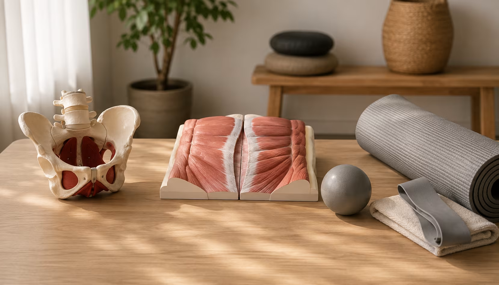
The rectus abdominis runs in two vertical bands from the pubic bone to the ribcage, joined down the middle by a strip of connective tissue called the linea alba. During pregnancy, this connective tissue stretches to accommodate the growing uterus. The two halves of the rectus separate — sometimes substantially. The condition has a clinical name: diastasis recti abdominis.
Some separation is universal. The question is how much, how long it persists, and whether it produces functional problems. The 2 finger-width threshold gets used a lot in postpartum literature, but the firmness of the tissue matters more than the gap width alone. A wider gap with healthy tension can be more functional than a narrower gap with soft, doming connective tissue.
The standard postnatal advice ("avoid crunches, do these specific exercises") has good evidence behind some of it and weaker evidence behind specific protocols. This piece walks through what actually helps, what doesn't, and how to think about diastasis without panicking over the social media wave of "is your ab gap normal?" content that's amplified in the past decade.
What Diastasis Recti Actually Is
The linea alba is connective tissue, not muscle. Composed of three sheets — the aponeuroses of the external oblique, internal oblique, and transverse abdominis — interwoven down the midline. During pregnancy, sustained pressure from the growing uterus, combined with hormonal changes that increase tissue laxity (relaxin), stretches the linea alba laterally. The two halves of the rectus abdominis end up further apart than they began.
The condition is universal in late pregnancy. Studies using ultrasound have found diastasis present in essentially 100% of pregnant women at 35 weeks gestation. The question is how the body recovers postpartum.
By 6 months postpartum, approximately 30–60% of women still show measurable diastasis. By 12 months, the number drops, but a substantial minority (around 25–35%) still have functional separation. Beyond 12 months, the resolution rate slows considerably; what remains at 18 months often requires intervention to change.
The functional issues that may follow:
- Reduced trunk stability and core strength
- Lower back pain (the rectus is part of the trunk's force coupling; weakness affects load distribution)
- Pelvic floor dysfunction (often comorbid)
- Abdominal aesthetic concerns (visible doming or bulge under tension)
- Increased risk of umbilical hernia at the most stretched section
The cosmetic and functional concerns can be separate. Significant cosmetic appearance doesn't always mean significant functional problem; significant functional problem can exist with modest visible appearance.
Self-Assessment
A reasonable self-check uses the following protocol:
- Lie on your back with knees bent and feet flat on the floor.
- Place fingertips along the midline at the level of the navel, perpendicular to the linea alba.
- Take a breath. Exhale. Lift your head slightly off the floor (think gentle chin tuck and lift, not full sit-up).
- Feel for the edges of the rectus muscle on either side of your fingers.
- Measure the gap in finger-widths.
- Repeat at 2 inches above the navel, at the navel, and 2 inches below.
Several findings inform interpretation:
Width
Reported in finger-widths historically; some clinicians use centimeters. Functionally:
- 2 finger-widths or less: typically considered functional, often improves further with time
- 2–3 finger-widths: mild diastasis, usually responds to focused work
- 3–4 finger-widths: moderate, may need physical therapy support
- 4+ finger-widths: significant, professional assessment warranted
Tension
Press into the gap. Firm, springy tension suggests functional connective tissue. Soft, deep, easily-depressible tissue suggests the linea alba hasn't restored normal tension. Width with tension can be more functional than narrower width without.
Doming
Watch the midline during the head lift. If a clear "tent" or ridge forms — the tissue bulging upward between the muscle bellies — this indicates inadequate core engagement during the movement. The doming is what to avoid.
Variation along length
The separation isn't usually uniform. Common pattern: smaller above navel, largest at and just below navel, narrowing again toward pubis. Test multiple locations.
For more accurate assessment, ultrasound is the gold standard. Some physical therapists who specialize in pelvic and abdominal health offer assessment that's more precise than self-testing.
Who's at Higher Risk
A few factors are associated with persistent diastasis:
Multiple pregnancies
The tissue stretches further with each pregnancy. Twins, triplets, and multiple previous pregnancies all increase risk.
Larger baby
The mechanical force scales with what the linea alba has had to accommodate.
Advanced maternal age
Connective tissue properties change with age; recovery capacity may be reduced.
Inadequate postpartum rest
Returning to high abdominal activity (heavy lifting, intense core work, jumping movements) in the first 6–8 weeks postpartum can prevent natural recovery.
Pre-existing connective tissue laxity
Ehlers-Danlos syndrome and other connective tissue conditions affect baseline tissue properties.
Genetic factors
Some women have stronger postpartum recovery than others for reasons not fully understood.
C-section vs vaginal delivery
The literature is mixed on whether C-section affects diastasis specifically. Recovery from the surgery itself adds healing demand that may slow general core recovery.
For men, the major risk factors are sustained intra-abdominal pressure: heavy lifting with poor technique, chronic obesity, chronic cough (asthma, smoking-related), and certain medical conditions producing ascites or abdominal distension.
What Resolves Naturally
The natural recovery story is more positive than alarming social media content suggests.
A 2021 study following women through the postpartum year found:
- At 6 weeks postpartum: roughly 60% had diastasis greater than 2.5 cm
- At 6 months postpartum: roughly 40% still had measurable diastasis
- At 12 months postpartum: roughly 30%
The drop from 6 weeks to 12 months reflects substantial natural healing. This happens in women with minimal targeted intervention — basic walking, daily activities, and time.
What seems to facilitate natural recovery:
- Adequate rest in the first 6 weeks
- Avoiding heavy lifting and high-pressure abdominal work early
- Side-lying when getting up rather than forceful crunching motions
- Gradual return to activity
- Adequate sleep (significantly affected for new parents — but real)
What hinders natural recovery:
- Aggressive return to crunches, planks, or heavy lifting in the first 6 weeks
- Chronic cough or chronic constipation (both produce repeated abdominal pressure)
- Significant breath-holding during lifting
- Poor pelvic floor coordination
For many women, the first six weeks of rest plus several months of progressive activity produces near-complete resolution without any specific protocol.
What the Exercises That Help Have in Common
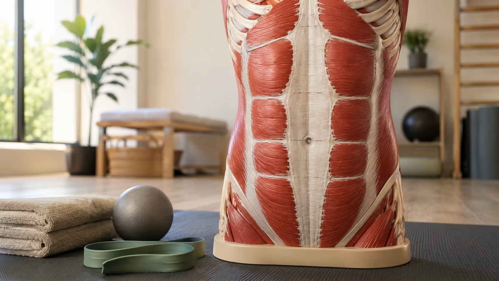
The exercises with evidence for diastasis recovery share several features:
They activate the transverse abdominis as a primary action
The TVA wraps around the abdomen like a corset. Its contraction draws the abdominal wall inward, which approximates the linea alba edges and produces appropriate tension across the connective tissue.
They train the deep core in connection with breathing
Exhalation naturally engages the TVA and pelvic floor. Inhalation expands the abdomen. The coordinated pattern is the goal, not isolated TVA contraction at random.
They avoid forward bulge or dome along the midline
If you can see the midline doming during an exercise, the exercise is doing the wrong thing for diastasis. Modify the position or load to eliminate the dome.
They use anti-extension and anti-rotation patterns more than flexion
Crunches train the rectus into flexion, which doesn't restore TVA function. Anti-extension exercises (dead bugs, bird dogs) and anti-rotation exercises (Pallof presses) train deep stability without producing the pattern that may have contributed to diastasis.
They build gradually from supine to upright to loaded positions
Early work happens lying down with minimal challenge. Progress involves adding the demand of gravity, then adding load. Skipping stages produces poor outcomes.
Specific Exercise Progressions
A reasonable progression for mild to moderate diastasis postpartum, starting at the 6–8 week mark and proceeding over months:
Phase 1: Foundation (Weeks 6–12 postpartum, or starting point for non-postpartum)
Belly breathing with TVA cuing
Lying on back, knees bent. Inhale and expand the belly outward (diaphragm descending). Exhale and gently draw the navel toward the spine — light engagement, no clenching. Coordinate the breath with the engagement. Repeat 8–10 times.
Pelvic floor breath coordination
Same position. As you exhale and engage TVA, also lift the pelvic floor (the action you'd use to stop urine flow, but gentle). Inhale and release both. Build coordination of the two.
Heel slides with stable trunk
Lying on back, knees bent. Maintain TVA engagement. Slide one heel out along the floor until the leg is straight, then return. The trunk shouldn't move; the midline shouldn't dome. 8–10 each leg.
Single-leg lifts with stable trunk
Similar position. Lift one knee toward the chest while maintaining trunk stability and TVA engagement. Lower without dropping the trunk. 8–10 each leg.
Phase 2: Progression (Weeks 12–24)
Dead bug
Lying on back, arms extended toward ceiling, knees bent 90 degrees over hips. Maintain TVA engagement. Slowly extend one arm and the opposite leg until both are nearly straight. Return. Alternate sides. 6–8 each side. Stop if midline domes.
Bird dog
On hands and knees, neutral spine. Extend opposite arm and leg simultaneously, maintaining trunk stability. Hold briefly, return. 6–8 each side.
Glute bridge with stable trunk
Lying on back, feet flat. Lift hips by engaging glutes, maintaining flat low back and TVA engagement. Lower under control. 10–12 reps.
Side plank from knees
Lying on side, propped on forearm, knees stacked. Lift hips so the body forms a straight line from knee to shoulder. Hold 10–20 seconds. Build to 30–45 seconds. Progress to side plank from feet.
Pallof press
Standing or half-kneeling, cable or band pulling laterally. Press the band out from your chest while resisting rotation. Hold and return. 8–10 each side.
Phase 3: Loaded Work (Months 6+)
Front-loaded squats and split squats
Goblet squats, front-rack split squats, Bulgarian split squats with light dumbbells. Maintain trunk stability.
Carries
Farmer's carries, suitcase carries (single side, anti-rotation challenge), front-loaded carries with goblet position.
Anti-extension press progressions
Wall push-ups → incline push-ups → modified push-ups → standard push-ups, with strict attention to midline stability.
Hip hinge progressions
Bodyweight RDLs → kettlebell RDLs → barbell deadlifts at light load, watching for midline integrity.
The progression is slower than people often want. Going from postpartum rest to barbell deadlifts in 3 months risks reversing recovery. The slower progression usually produces better long-term outcomes.
What to Avoid (and Why)
Several common exercises that may worsen diastasis:
Crunches and sit-ups
Repeatedly load the rectus abdominis into flexion against the connective tissue. Reinforce the forward pressure pattern.
Russian twists and oblique crunches
Same forward pressure plus rotation that adds shear stress to the linea alba.
Full planks (high or low) before adequate TVA function
Until you can hold a plank without midline doming, the exercise is loading the connective tissue in ways that don't help.
Heavy lifting with breath-holding
Valsalva maneuver under load creates intra-abdominal pressure that may stretch the linea alba further.
Jumping movements with poor core engagement
High-impact landings spike intra-abdominal pressure repeatedly.
Excessive forward bending without core engagement
Pulling weight from the floor in poor positions, repetitive forward folds with poor brace.
V-sits, leg lifts in long lever (legs straight). High abdominal pressure from below.
These aren't permanently forbidden. They're inappropriate during the rehabilitation phase. Once diastasis has resolved and core function is restored, normal activity returns. The timeline is months, not weeks, for moderate cases.
When to See a Specialist
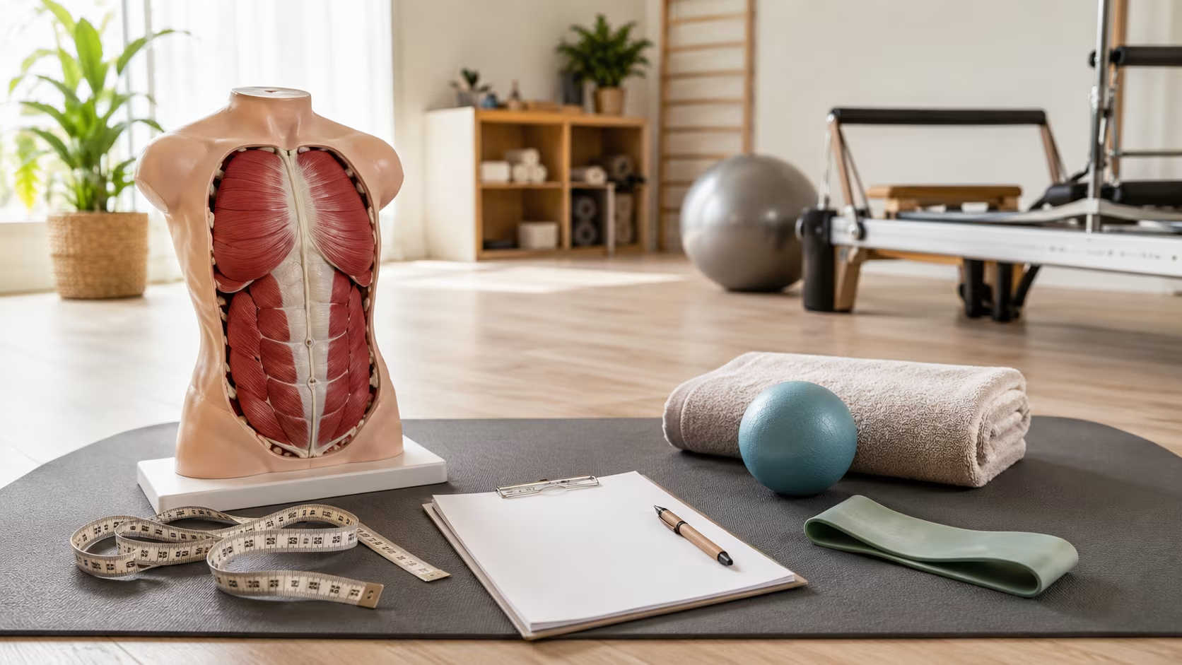
A few markers that suggest professional evaluation:
- Diastasis greater than 3 finger-widths persisting past 3 months postpartum
- Significant pelvic floor symptoms (urinary leakage, pelvic heaviness, painful intercourse)
- Visible umbilical hernia (bulging through the navel area)
- Diastasis with significant back pain that doesn't respond to general core work
- No improvement after 12 weeks of focused work
- Doming or bulging that won't go away even at rest
The specialist to seek is a physical therapist with specific training in pelvic health or postpartum rehabilitation. The credential most commonly listed is PT, DPT with additional training in pelvic floor or women's health. Some programs certify additional specialization (CAPP-OB, BCB-PMD).
These specialists can:
- Provide accurate ultrasound assessment
- Identify coexisting pelvic floor issues
- Design progression specific to your presentation
- Address overlapping issues (pelvic floor weakness, postural patterns, breathing dysfunction)
- Decide if surgical consultation is appropriate
In many countries, pelvic PT is covered by insurance with appropriate referral. In others, it requires out-of-pocket payment.
The Surgery Question
For diastasis that doesn't respond to conservative care, surgical repair is an option. Standard approach: abdominoplasty (tummy tuck) with plication of the linea alba — the surgeon sews the two halves of the rectus back together along the midline.
Indications for surgical consideration:
- Persistent diastasis greater than 5 cm despite extensive conservative care
- Functional impairment (significant ongoing back pain, hernia formation)
- Cosmetic concerns that significantly affect quality of life
- Family planning complete (the surgery would be undone by subsequent pregnancy)
Recovery from abdominoplasty is substantial — typically 4–8 weeks of significantly limited activity, with full return to athletic activity taking 3–6 months. The surgery is cosmetic in many insurance frameworks and often not covered, though some plans cover repair when functional impairment is documented.
For most women with diastasis, conservative care produces adequate resolution. Surgery is for the persistent cases that haven't responded to focused rehabilitation.
Sources
Sperstad, J. B., et al. (2016). Diastasis recti abdominis during pregnancy and 12 months after childbirth: prevalence, risk factors and report of lumbopelvic pain. British Journal of Sports Medicine, 50(17), 1092–1096.
Benjamin, D. R., van de Water, A. T., & Peiris, C. L. (2014). Effects of exercise on diastasis of the rectus abdominis muscle in the antenatal and postnatal periods: a systematic review. Physiotherapy, 100(1), 1–8.
Mota, P., et al. (2015). Prevalence and risk factors of diastasis recti abdominis from late pregnancy to 6 months postpartum, and relationship with lumbo-pelvic pain. Manual Therapy, 20(1), 200–205.
Litos, K. (2014). Progressive therapeutic exercise program for successful treatment of a postpartum woman with a severe diastasis recti abdominis. Journal of Women's Health Physical Therapy, 38(2), 58–73.
Hills, N. F., Graham, R. B., & McLean, L. (2018). Comparison of trunk muscle function between women with and without diastasis recti abdominis at 1 year postpartum. Physical Therapy, 98(10), 891–901.
Gluppe, S. L., et al. (2018). Effect of a postpartum training program on the prevalence of diastasis recti abdominis in postpartum primiparous women: a randomized controlled trial. Physical Therapy, 98(4), 260–268.
Frequently Asked Questions
How do I check for diastasis recti?
Lie on your back, knees bent, feet flat. Place fingers across your midline, just above the navel. Lift your head slightly. Feel for the gap between the two halves of the rectus abdominis. Measure in finger-widths. Test above, at, and below the navel — the separation can vary along the linea alba.
How wide is a 'normal' diastasis after pregnancy?
Some separation is universal during and shortly after pregnancy. By 6–8 weeks postpartum, a gap of 2 finger-widths or less is considered functionally normal. Wider gaps may benefit from focused work. The width matters less than the firmness of the connective tissue — a 2-finger gap with good tension is more functional than a 1-finger gap with significant doming.
Will my diastasis ever close completely?
Many resolve to functional baseline within 8–12 weeks postpartum with adequate rest and gentle deep core work. Some don't fully close to a hairline gap but become functionally fine. Persistent significant separation past 6 months postpartum may benefit from physical therapy specifically trained in this area. Surgery (abdominoplasty with diastasis repair) is reserved for cases that don't respond to conservative care.
Can I do regular ab exercises with diastasis?
Crunches, sit-ups, and Russian twists usually make diastasis worse, not better. They reinforce the same forward force on the linea alba that pregnancy already produced. The work that helps involves activating the deep transverse abdominis and obliques without creating dome or bulge along the midline.
Is diastasis recti only a postpartum issue?
No. Men can develop it from chronic intra-abdominal pressure (heavy lifting with poor bracing, sustained obesity, chronic cough). Postpartum women are the largest group, but the condition isn't gender-specific. The mechanism is the same — sustained outward pressure stretches the linea alba beyond its normal recovery capacity.





0 Comments
Leave a comment
Your email won't be published. Required fields are marked with *.